Let’s delve into the fascinating realm of nuclear medicine, an advanced branch of medical science that employs radioactive substances to diagnose and treat a wide array of ailments.
In this article, we will explore the intricacies of nuclear medicine and shed light on its functioning.
You might have seen patients undergoing radiation therapy for cancer and doctors ordering PET scans to diagnose patients.
These are but small parts of the medical specialty called “Nuclear Medicine”.
Nuclear medicine uses radioactive substances to image the body and treat disease.
It looks at both the physiology (functioning) and the anatomy (constitution – what is it made of) of the body in establishing diagnosis and treatment.
What is “Diagnosis”?
The term “diagnosis” refers to the process of identifying and determining the nature or cause of a medical condition or disease through examination, evaluation, and analysis of symptoms, medical history, and diagnostic tests.
It involves the identification and classification of the specific ailment or disorder afflicting an individual, enabling healthcare professionals to develop an appropriate treatment plan.
What is Nuclear Medicine?
Nuclear medicine is a specialized field of medicine that utilizes small amounts of radioactive materials, known as “radiotracers”, to examine and diagnose medical conditions.
Unlike other diagnostic techniques, such as X-rays or CT scans, nuclear medicine focuses on evaluating the functional processes within the body at a cellular and molecular level.
Imaging And Diagnostics in Nuclear Medicine
The human body is opaque and doctors can’t look into the human body without the help of modern imaging techniques.
Previously, exploratory surgery was prevalent for internal examinations. Yes! Surgeons used to cut up the body just to explore if they could observe anything wrong. Would you have liked that? Me? NEVER!
However, contemporary medicine offers an extensive range of non-invasive procedures.
These include X-rays, MRI scanners, CAT scans, ultrasound, and more.
Each technique possesses distinct merits and drawbacks, rendering them suitable for specific conditions and body areas.
Nuclear medicine imaging techniques give doctors another way to look inside the human body.
The techniques combine the use of computers, detectors, and radioactive substances. These techniques include:
Nuclear imaging is useful for detecting:
The use of any specific test, or combination of tests, depends upon the patient’s symptoms and the disease being diagnosed.
Positron Emission Tomography (PET)
PET produces images of the body by detecting the radiation emitted from radioactive substances.
These substances are injected into the body, and are usually tagged with a radioactive atom, such as Carbon-11, Fluorine-18, Oxygen-15, or Nitrogen-13, that have a short decay time.
These radioactive atoms are formed by bombarding normal chemicals with neutrons to create short-lived radioactive isotopes.
PET detects the gamma rays given off at the site where a positron emitted from the radioactive substance collides with an electron in the tissue
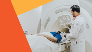
In a PET scan, the patient is injected with a radioactive substance and placed on a flat table that moves in increments through a “doughnut” shaped housing.
This housing contains the circular gamma ray detector array, which has a series of scintillation crystals, each connected to a photomultiplier tube.
The crystals convert the gamma rays, emitted from the patient, to photons of light, and the photomultiplier tubes convert and amplify the photons to electrical signals.
These electrical signals are then processed by the computer to generate images.
The table is then moved, and the process is repeated, resulting in a series of thin slice images of the body over the region of interest (e.g. brain, breast, liver).
These thin slice images can be assembled into a three dimensional representation of the patient’s body.
PET provides images of blood flow or other biochemical functions, depending upon the type of molecule that is radioactively tagged.
For example, PET can show images of glucose metabolism in the brain, or rapid changes in activity in various areas of the body.
However, there are few PET centers in the country because they must be located near a particle accelerator device that produces the short-lived radioisotopes used in the technique.
Here are some of the best PET centers in the country.
SPECT, Cardiovascular Imaging and Bone Scanning
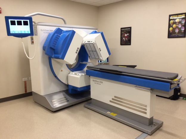
SPECT is a technique similar to PET. But the radioactive substances used in SPECT (Xenon-133, Technetium-99, Iodine-123) have longer decay times than those used in PET, and emit single instead of double gamma rays.
SPECT can provide information about blood flow and the distribution of radioactive substances in the body.
Its images have less sensitivity and are less detailed than PET images, but the SPECT technique is less expensive than PET.
Cardiovascular imaging techniques use radioactive substances to chart the flow of blood through the heart and blood vessels.
One example of a cardiovascular imaging technique is a stress thallium test, in which the patient is injected with a radioactive thallium compound, exercised on a treadmill, and imaged with a gamma ray camera.
After a period of rest, the study is repeated without the exercise.
The images before and after exercising are compared to reveal changes in blood flow to the working heart.
These techniques are useful in detecting blocked arteries or arterioles in the heart and other tissues.
Bone scanning detects radiation from a radioactive substance (technetium-pp methyl diphosphate) that, when injected into the body, collects in bone tissue, as bone tissue is good at accumulating phosphorus compounds.
The substance accumulates in areas of high metabolic activity, and so the image produced shows “bright spots” of high activity and “dark spots” of low activity.
Bone scanning is useful for detecting tumors, which generally have very high metabolic activity.
Treatment in Nuclear Medicine
In nuclear medicine imaging tests, injected radioactive substances pose little harm to the body when injected in small doses.
However, note that radiation kills live cells and the choice is between how useful it is to the small loss that will occur.
If the benefits are more than the damage causes, doctors go ahead with these scans.
Furthermore excessive radiation testing increases the risk of cancer due to radiation and in medically negligent cases where high doses are negligently given, a radiation burn (internal or external) and many other issues
Radioisotopes that decay quickly, emit lower radiation levels than X-rays or CT scans, and are eliminated through urine or bowel movements.
However, specific cells are highly affected by ionizing radiation, including alpha, beta, gamma, and X-rays and hence pose some amount of risk. Hence, excessive testing must be avoided. Sometimes “symptoms” reveal more than “tests” – a good doctor is hence invaluable.
There’s a saying in India – वैद्यो नारायणो हरि: १. Which means doctors are equal to the supreme narayana – The god.
Cells with rapid multiplication rates are more susceptible to radiation damage due to two key factors:
Fast-multiplying cells have limited time for DNA repair before division, increasing the likelihood of self-destruction when exposed to nuclear radiation.
As cancer often involves rapidly dividing cells, radiation therapy can be utilized for treatment.
For instance, radioactive wires or vials are placed near or around the tumor. In cases of deep or inoperable tumors, high-intensity X-rays are focused on the affected area.
However, this form of treatment can also impact normal cells that are in the replication stage.
Cells in the hair, stomach and intestinal lining, skin, and blood cells are highly sensitive to radiation due to their rapid reproduction. Consequently, hair loss and nausea are common side effects experienced by individuals undergoing cancer treatment.
Furthermore, nuclear materials are employed to produce radioactive tracers injected into the bloodstream.
One type of tracer allows visualization of the blood vessel structure, facilitating easy detection of clots and abnormalities.
Certain organs concentrate specific chemicals. For example, the thyroid gland concentrates iodine, enabling the detection of certain thyroid tumors through the injection of radioactive iodine into the bloodstream.
Similarly, cancerous tumors concentrate phosphates. By injecting the radioactive phosphorus-32 isotope into the bloodstream, tumors can be detected by their increased radioactivity.
Nuclear technology when used in a non destructive way will open a new world of possibilities that can usher humanity into a new era of technology enabled life.
However, it’s important to monitor and be cautious with the usage of nuclear and related technology due to safety concerns.