Understanding MRI Machines: Invention, Function & Insights

MRI or magnetic resonance imaging is a commonly prescribed test in the healthcare sector to diagnose health problems.
Have you ever looked at the jumbo sized MRI machines and wondered how it works?
Delve into this comprehensive guide to unravel the mystery behind this remarkable medical imaging technology. From its invention to the fundamental principles governing its operation, we’ll cover everything you need to know about MRI machines.
The Invention and History
When you have a health issue or traumatic injury that needs to be treated, before the medical imaging techniques were invented, doctors had to cut you open to find what the issue was.
Cutting someone open to find an issue and then later fixing it is not an ideal or even a possible solution.
Scientists and physicians have discovered and invented methods to scan the body for issues without cutting open for years.
Dr. Raymond Damadian, a physician and scientist, toiled for years trying to produce a machine that could noninvasively scan the body with the use of magnets.
Along with some graduate students, he constructed a superconducting magnet and fashioned a coil of antenna wires.
Since no one wanted to be the first one in this contraption, Damadian volunteered to be the first patient.
When he climbed in, however, nothing happened.
Damadian was looking at years wasted on a failed invention, but one of his colleagues bravely suggested that he might be too big for the machine.
A svelte graduate student volunteered to give it a try, and on July 3, 1977, the first MRI exam was performed on a human being.
It took almost five hours to produce one image, and that original machine, named the “Indomitable,” is now owned by the Smithsonian Institution.
In just a few decades, the use of magnetic resonance imaging (MRI) scanners has grown tremendously.
Doctors may order MRI scans to help diagnose multiple sclerosis, brain tumors, torn ligaments, tendonitis, cancer and strokes, to name just a few.
An MRI scan is the best way to see inside the human body without cutting it open.
That may be little comfort to you when you’re getting ready for an MRI exam.
You’re stripped of your jewelry and credit cards and asked detailed questions about all the metallic instruments you might have inside of you.
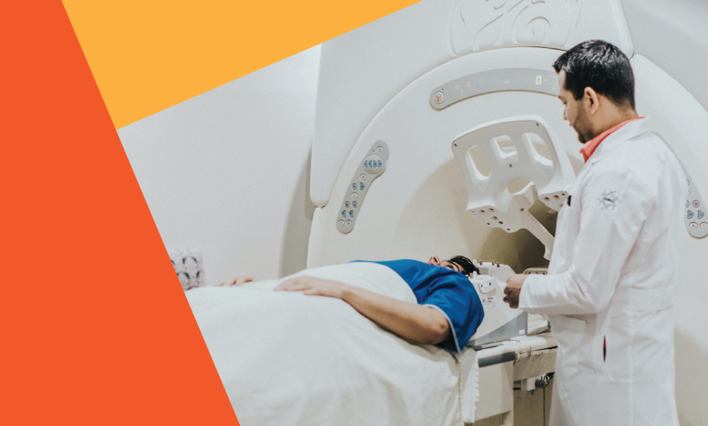
You’re then put on a tiny slab and pushed into a hole with a lot of plastic tubing and machines in it.
When the MRI starts, you must lay still and are blasted with loud noises with different sound patterns.
And with each minute, you can’t help but wonder what’s happening to your body while it’s in this machine.
Could it really be that this ordeal is truly better than another imaging technique, such as an X-ray or a CAT scan? What has Raymond Damadian wrought?
What Is An MRI?
An MRI (magnetic resonance imaging) scan is a painless test that produces very clear images of the organs and structures inside your body.
MRI uses a large magnet, radio waves and a computer to produce these detailed images. It doesn’t use X-ray or radiation.
Because MRI doesn’t use X-rays or other radiation, it’s the imaging test of choice when people will need frequent imaging for diagnosis or treatment monitoring, especially of their brain.
Working Of An MRI Machine
Modern MRI machines h=come in different sizes, power and shapes with magnets being the main component used.
However the basic design and working of an MRI machine is the same since invention and the patient is pushed inside a long tube.
The MRI tube is only about 60 centimeters in diameter which is a limitation for overly obese patients.
The biggest and most important component of an MRI system is the magnet in it.
There is a horizontal tube where the patient is placed and has magnets running through the tube from the front to back.
This tube is known as the bore. But this isn’t just any ordinary magnet that you can buy.
The magnetic systems here are strong here, one capable of producing a large, stable magnetic field.
The strength of a magnet in an MRI system is rated using a unit of measure known as a tesla.
Another unit of measure commonly used with magnets is the gauss where 1 tesla is 10,000 gauss.
The magnets in use today in MRI systems create a magnetic field of 1.5-tesla to 3.0-tesla, or 15,000 to 30,000 gauss.
There are also MRI machines which can create a magnetic field of up to 7 tesla too, However they are not widely used.
Note that the Earth’s magnetic field measures 0.5 gauss, you can see how powerful these magnets are.
Most MRI systems use a superconducting magnet, which consists of many coils or windings of wire through which a current of electricity is passed, creating a magnetic field of up to 3.0 tesla.
Maintaining such a large magnetic field requires a good deal of energy, which is accomplished by superconductivity, or reducing the resistance in the wires to almost zero.
To do this, the wires are continually bathed in liquid helium at 452.4 degrees below zero Fahrenheit or -269.1 degrees Celsius.
These cold wires are insulated by vacuum so as not to cool other areas or the patient inside.
While superconductive magnets are expensive, the strong magnetic field allows for the highest-quality imaging, and superconductivity keeps the system economical to operate.
Since the Magnetic field is activated only when the MRI machine is powered and switched on, it’s easier to maintain.
Using an electromagnet has another advantage here over a normal magnet because a normal powerful magnet will lose it’s power over time and needs frequent replacement.
Two other magnets are used in MRI systems to a much lesser extent.
Resistive magnets are structurally like superconducting magnets, but they lack the liquid helium.
This difference means they require a huge amount of electricity, making it prohibitively expensive to operate above a 0.3 tesla level.
Permanent magnets have a constant magnetic field, but they’re so heavy that it would be difficult to construct one that could sustain a large magnetic field.
There are also three gradient magnets inside the MRI machine.
These magnets are much lower strength compared to the main magnetic field; they may range in strength from 180 gauss to 270 gauss.
While the main magnet creates an intense, stable magnetic field around the patient, the gradient magnets create a variable field, which allows different parts of the body to be scanned.
Another part of the MRI system is a set of coils that transmit radiofrequency waves into the patient’s body.
There are different coils for different parts of the body: knees, shoulders, wrists, heads, necks and so on.
These coils usually conform to the contour of the body part being imaged, or at least reside very close to it during the exam.
Other parts of the machine include a very powerful computer system and a patient table, which slides the patient into the bore.
Whether the patient goes in head or feet first is determined by what part of the body needs examining.
Once the body part to be scanned is in the exact center, or isocenter, of the magnetic field, the scan can begin.
Advancements in MRI technology
MRI machines are continuously evolving to make it more patient-friendly.
For example, many claustrophobic people simply can’t stand the cramped confines, and the bore may not accommodate obese people.
There are more open scanners, which allow for greater space, but these machines have weaker magnetic fields,
meaning it may be easier to miss abnormal tissue.
Very small scanners for imaging specific body parts are also being developed. Other advancements are being made in the field of MRI.
Functional MRI (fMRI), for example, creates brain maps of nerve cell activity second by second and is helping researchers better understand how the brain works.
Magnetic resonance angiography (MRA) creates images of flowing blood, arteries and veins in virtually any part of the body.
The principle and How It Scans Your Body
The human body is made of billions of atoms and various chemicals like hydrogen, oxygen to name a few.
For the purposes of an MRI scan, we’re only concerned with the hydrogen atoms in your body, which is abundant since the body is mostly made up of water and fat.
These atoms are randomly spinning on their axis.
All of the atoms are going in various directions, but when placed in a magnetic field, the atoms line up in the direction of the field.
These hydrogen atoms have a strong magnetic moment, which means that in a magnetic field, they line up in the direction of the field.
Since the magnetic field runs straight down the center of the machine, the hydrogen protons line up so that they’re pointing to either the patient’s feet or the head.
About half go each way, so that the vast majority of the protons cancel each other out — that is, for each atom lined up toward the feet, one is lined up toward the head.
Only a couple of protons out of every million aren’t canceled out.
This doesn’t sound like much, but the sheer number of hydrogen atoms in the body is enough to create extremely detailed images.
It’s these unmatched atoms that we’re concerned with now.
Next, the MRI machine applies a radio frequency (RF) pulse that is specific only to hydrogen.
The system directs the pulse toward the area of the body we want to examine.
When the pulse is applied, the unmatched protons absorb the energy and spin again in a different direction.
This is the “resonance” part of MRI.
The RF pulse forces them to spin at a particular frequency, in a particular direction.
The specific frequency of resonance is called the Larmour frequency and is calculated based on the particular tissue being imaged and the strength of the main magnetic field.
At approximately the same time, the three gradient magnets jump into the act.
They are arranged in such a manner inside the main magnet that when they’re turned on and off rapidly in a specific patterns, they alter the main magnetic field on a local level.
What this means is that we can pick exactly which area we want a picture of; this area is referred to as the “slice.”
Think of a loaf of bread with slices as thin as a few millimeters — the slices in MRI are that precise.
Slices can be taken of any part of the body in any direction, giving doctors a huge advantage over any other imaging modality.
That also means that you don’t have to move for the machine to get an image from a different direction — the machine can manipulate everything with the gradient magnets.
But the machine makes a tremendous amount of noise during a scan, which sounds like a continual rapid hammering.
That’s due to the rising electrical current in the wires of the gradient magnets being opposed by the main magnetic field.
The stronger the main field, the louder the gradient noise. In most MRI centers, you can bring a music player to drown out the racket, and patients are given earplugs.
When the RF pulse is turned off, the hydrogen protons slowly return to their natural alignment.
within the magnetic field and release the energy absorbed from the RF pulses.
When they do this, they give off a signal that the coils pick up and send to the computer system.
But how is this signal converted into a picture that means anything?
How Are Readable MRI Images Generated?
The MRI scanner can pick out a very small point inside the patient’s body and ask it, essentially, “What type of tissue are you?”
The system goes through the patient’s body point by point, building up a map of tissue types.
It then integrates all of this information to create 2-D images or 3-D models with a mathematical formula known as the Fourier transform.
The computer receives the signal from the spinning protons as mathematical data; the data is converted into a picture. That’s the “imaging” part of MRI.
The MRI system uses injectable contrast, or dyes, to alter the local magnetic field in the tissue being examined.
Normal and abnormal tissue respond differently to this slight alteration, giving us differing signals.
These signals are transferred to the images; an MRI system can display more 250 shades of gray to depict the varying tissue.
The images allow doctors to visualize different types of tissue abnormalities better than they could without the contrast.
We know that when we do “A,” normal tissue will look like “B” — if it doesn’t, there might be an abnormality.
An X-ray is very effective for showing doctors a broken bone, but if they want a look at a patient’s soft tissue, including organs, ligaments and the circulatory system, then they’ll likely want an MRI.
And, as we mentioned earlier, another major advantage of MRI is its ability to image in any plane.
Computer tomography (CT), for example, is limited to one plane, the axial plane (in the loaf-of-bread analogy, the axial plane would be how a loaf of bread is normally sliced).
An MRI system can create axial images as well as sagitall (slicing the bread side-to-side lengthwise) and coronal (think of the layers in a layer cake) images, or any degree in between, without the patient ever moving.
But for these high-quality images, the patient shouldnt move at all, staying still and relaxed throughout the procedure.
MRI scans require patients to hold still for 20 to 90 minutes or more.
Even very slight movement of the part being scanned can cause distorted images that will have to be repeated.
And there’s a high cost to this kind of quality; MRI systems are very expensive to purchase, and therefore the exams are also very expensive.
Safety Concerns Of An MRI Scan
An MRI scan is generally safe and poses almost no risk to the average person when appropriate safety guidelines are followed.
Even though all your atoms are mixed about during a scan, once you’re out of the magnetic field, your body and its chemistry return to normal.
The strong magnetic field the MRI machines emit is not harmful to you, but it may cause implanted medical devices to malfunction or distort the images.
The fact that MRI systems don’t use ionizing radiation, as other imaging devices do, is a comfort to many patient.
Also the MRI contrast materials if used have a very low incidence of side effects.
Most often doctors prefer not to image pregnant women, due to limited research of the biological effects of magnetic fields on a developing fetus.
The decision of whether or not to scan a pregnant patient is made on a case-by-case basis with consultation between the MRI radiologist and the patient’s obstetrician.
However, the MRI suite can be a very dangerous place if strict precautions are not observed.
Credit cards or anything else with magnetic encoding will be erased. Metal objects can become dangerous projectiles if they are taken into the scan room.
For example, paperclips, pens, keys, scissors, jewelry, stethoscopes and any other small objects can be pulled out of pockets and off the body without warning, at which point they fly toward the opening of the magnet at very high speeds.
Big objects pose a risk, too — mop buckets, vacuum cleaners, IV poles, patient stretchers, heart monitors and countless other objects have all been pulled into the magnetic fields of the MRI.
In 2058 VS, a young boy undergoing a scan was killed when an oxygen tank was pulled into the magnetic bore.
Once, a pistol flew out of a policeman’s holster, the force causing the gun to fire. No one was injured.
To ensure safety, patients and support staff should be thoroughly screened for metal objects prior to entering the scan room.
Often, however, patients have implants inside them that make it very dangerous for them to be in the presence of a strong magnetic field.
These include:
- Metallic fragments in the eye, which are very dangerous as moving these fragments could cause eye damage or blindness
- Pacemakers, which may malfunction during a scan or even near the machine
- Aneurysm clips in the brain, which could tear the very artery they were placed on to repair if the magnet moves them
- Dental implants, if magnetic
Most modern surgical implants, including staples, artificial joints and stents are made of non-magnetic materials, and even if they’re not, they may be approved for scanning.
But let your doctor know, as some orthopedic hardware in the area of a scan can cause distortions in the image.
When Are MRI Scans Prescribed?
Healthcare providers use magnetic resonance imaging (MRI) to help diagnose or monitor the treatment for many different conditions.
There are also different types of MRIs based on which area of your body your provider wants to examine.
Brain and spinal cord MRIs can help evaluate and diagnose the following conditions:
- Brain aneurysms.
- Brain tumors and spinal tumors.
- Brain and spine injuries from trauma.
- Compression or inflammation of spinal cord and nerves (pinched nerve).
- Multiple sclerosis (MS).
- Spinal cord conditions.
- Spine anatomy and alignment.
- Stroke.
Providers use cardiac (heart) MRIs for several reasons, including:
- To evaluate the anatomy and function of your heart chambers, heart valves, size of and blood flow through major vessels and the surrounding structures.
- To diagnose cardiovascular conditions, such as tumors, infections and inflammatory conditions.
- To evaluate the effects of coronary artery disease, such as limited blood flow to your heart muscle and scarring within your heart muscle after a heart attack.
- To evaluate the anatomy and function of the heart and blood vessels in children and adults with congenital heart disease.
Body MRIs can evaluate structures and diagnose several conditions, including:
- Tumors in your chest, abdomen or pelvis.
- Liver diseases, such as cirrhosis, and issues with your bile ducts and pancreas.
- Inflammatory bowel disease, such as Crohn’s disease and ulcerative colitis.
- Malformations of blood vessels and inflammation of the vessels (vasculitis).
- A developing fetus in your uterus.
MRIs of bones and joints can help evaluate:
- Bone infections (osteomyelitis).
- Bone tumors.
- Disk abnormalities in your spine.
- Joint issues caused by injuries.
Healthcare providers sometimes use breast MRIs with mammography to detect breast cancer, especially in people who have dense breast tissue or who might be at high risk of breast cancer.


