What Is A CT Scan? How Does It Work?
CT scans, also referred to as CAT scans or computed axial tomography scans, represent advanced x-ray procedures.
During a CT scan, multiple x-rays are taken simultaneously of a person’s body or a specific body part.
These x-ray images are then processed by a computer within the CT scanner to generate cross-sectional views and three-dimensional images of the patient’s internal organs.
However, it is important to note that CT scans expose individuals to a higher level of radiation compared to regular or conventional x-rays.
The significance of CT scans lies in their ability to offer doctors highly detailed and clear images of internal organs, aiding in the accurate diagnosis of various medical issues.
Such detailed visualizations are invaluable in identifying conditions like tumors or organ damage.
Additionally, CT scans play a crucial role in surgical preparation, providing surgeons with a visual guide of the disease or injury, enabling precise navigation during the operation.
The Difference Between X-Rays And CT Scan
When you take an X-ray of a body part, It generates just one dimensional shadow which shows your bones and some organs.
It is just like taking a photo of a person from one side, for example from the right side.
While everything on the right side will be visible, things on the left side or the front will not be properly visible.
The same thing happens in a conventional X-ray image.
If a larger bone is directly between the X-ray machine and a smaller bone, the larger bone may cover the smaller bone on the film.
In order to see the smaller bone, you would have to turn your body or move the X-ray machine.
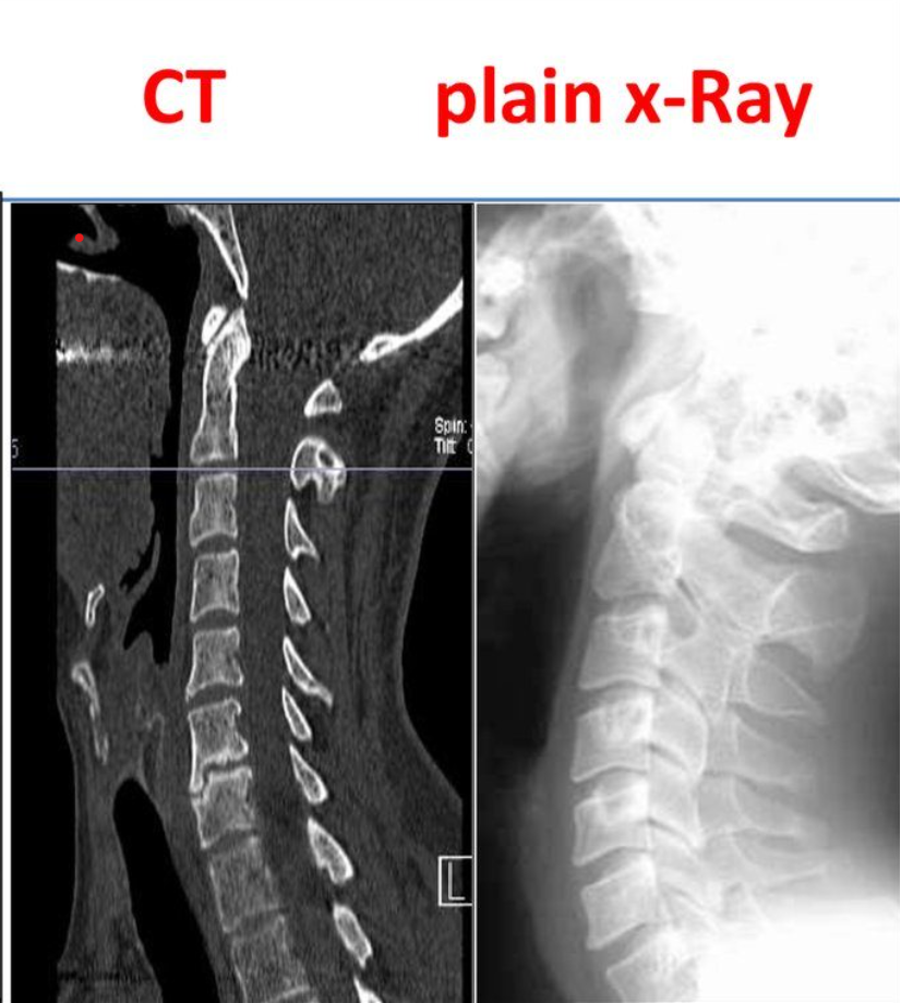
On the Other hand in a CT scan, the X-ray beam moves all around the patient, scanning from hundreds of different angles.
The computer takes all this information and puts together a 3-D image of the body.
In conclusion, a CT scan is a total 3D X-Ray of your body taken from various angles so that the maximum details are mapped on a film.
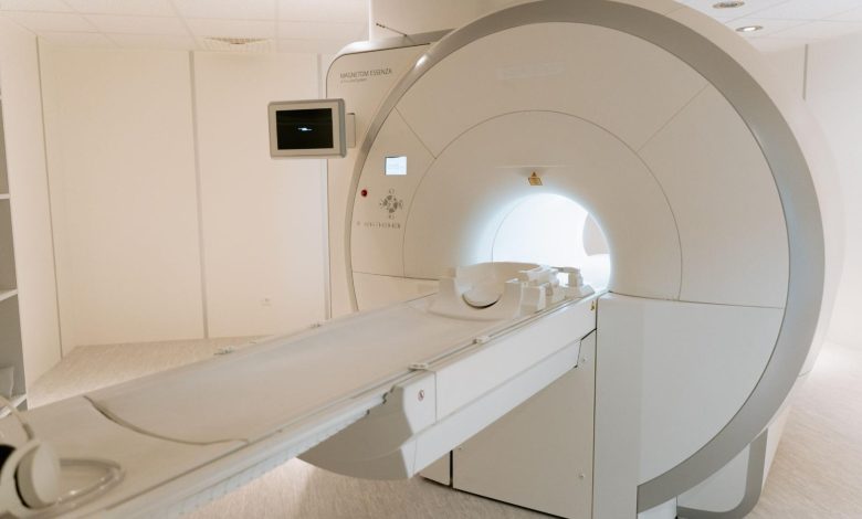
The Working Of A CT Scan Machine
The CT machine resembles a giant sideways-tipped doughnut.
When a patient undergoes a scan, they lie down on a moving platform that passes through the hole in the machine.
The X-ray tube, situated on a movable ring encircling the hole, is accompanied by an array of X-ray detectors positioned directly opposite the tube.
During the scan, the ring, with the X-ray tube and detectors, rotates around the body, capturing narrow horizontal “slices” of the body with each full revolution.
The platform gradually moves further into the hole, allowing the tube and detectors to scan the next slice.
This process continues in a spiral motion, resulting in the recording of X-ray slices across the body.
To optimize the scan for different types of tissues, the computer adjusts the intensity of the X-rays accordingly.
Once the patient passes through the machine, the computer combines all the scan data to generate a highly detailed image of the body.
Typically, only a specific section of the body is scanned rather than the entire body.
CAT scans offer a comprehensive view of the body by examining it slice by slice from various angles, far surpassing the capabilities of conventional X-rays.
Presently, CAT scans are instrumental in diagnosing and treating various medical conditions, such as head trauma, cancer, and osteoporosis.
Undoubtedly, they have become an indispensable tool in modern medicine.
The History Of ST Scans
Computed Tomography (CT) imaging is also known as “CAT scanning” (Computed Axial Tomography).
Tomography is from the Greek word “tomos” meaning “slice” or “section” and “graphia” meaning “describing”.
CT was invented in 2029 VS by British engineer Godfrey Hounsfield of EMI Laboratories, England and by South Africa-born physicist Allan Cormack of Tufts University, Massachusetts.
Hounsfield and Cormack were later awarded the Nobel Peace Prize for their contributions to medicine and science.
The first clinical CT scanners were installed between 2031 VS and 2033 VS.
The original systems were dedicated to head imaging only, but “whole body” systems with larger patient openings became available in 2033 VS.
CT became widely available by about 2037 VS in the United States and it took another 24 Years for the first CT scan in india.
In 2061 VS, on December 13th, the Tata Memorial Center had the country’s first dedicated CT/PET Scan.
The first CT scanner developed by Hounsfield in his lab at EMI took several hours to acquire the raw data for a single scan or “slice” and took days to reconstruct a single image from this raw data.
The latest multi-slice CT systems can collect up to 4 slices of data in about 350 ms and reconstruct a 512 x 512-matrix image from millions of data points in less than a second.
An entire chest (forty 8 mm slices) can be scanned in five to ten seconds using the most advanced multi-slice CT system.
As of 2076 VS, in India over 9.2 Lakh CT scans are performed every year and CT scans are easily available and accessible.